In the next decade or so, arthritis, especially knee arthritis, is expected to emerge as the fourth most common cause of physical disability in India. Our country is seeing an epidemic of arthritis, and its incidence in the Indian population is believed to be much higher than what is found in Western nations. It will be difficult for our country to tackle this colossal healthcare burden due to a shortage of healthcare infrastructure and rehabilitation specialists.
A significant reason for the rapid rise in knee arthritis is that life expectancy in India has doubled since Independence, leading to a massive pool of ageing population suffering from wear and tear of the knees.
The most common arthritis seen in India is age-related degenerative arthritis, which involves degeneration (wear and tear) of cartilage and can affect any joint such as the knee. In Indian women, the average age for the onset of knee problems is 50 years while in Indian males, it is 60 years. The reasons for early onset in females include obesity and poor nutrition. As many as 90 per cent of Indian women are deficient in vitamin D, which is critical in controlling bone metabolism. Its absence in the body directly or indirectly affects the knee. The lack of structured strength and mobility enhancement programmes in most people’s lives makes them prone to degenerative arthritic conditions.
Arthritis is a general term that applies to joint inflammation. The two primary forms are osteoarthritis (OA) and rheumatoid arthritis (RA). OA results from the degeneration of synovial fluid and generally progresses into a loss of articular cartilage, which typically presents itself as localised joint pain and a reduction of range of motion (ROM). This is the main cause of knee pain in middle-aged and elderly subjects. RA is a chronic autoimmune disease that results in inflammation of the synovium, leading to long-term joint pain, chronic pain and loss of function and mobility.
The scene in India
In India, almost 1.3 crore people suffer from RA and the disease is not restricted to the older generation. Most people who are affected by RA belong to the younger age group — between 20 and 30 years. It is also more rampant among women.
What is autoimmune arthritis?
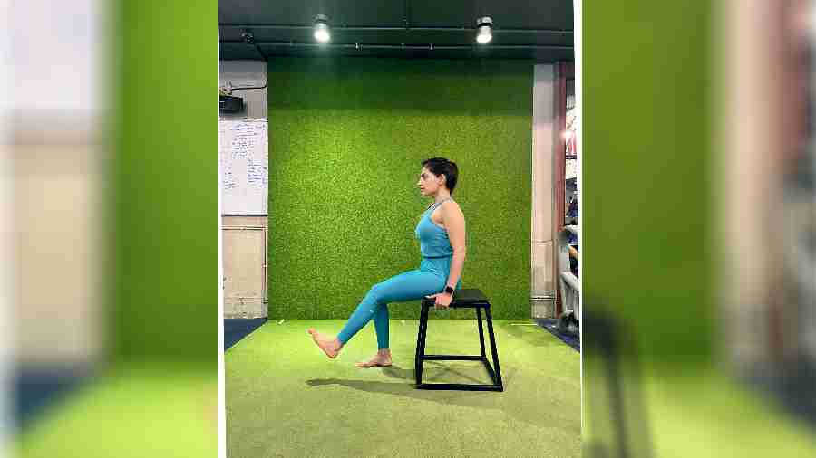
Arthritis patients should always perform an adequate warmup session
Auto-immune diseases are caused by the malfunctioning of our immune system. This means that sometimes our immune system (whose primary function is to protect us from germs, viruses, infections and so on) actually starts attacking our bodies instead of protecting us. Medical scientists are of the opinion that a malfunctioning or “leaky” gut may be the root cause of all autoimmune diseases.
The following are some forms of autoimmune arthritis:
- Rheumatoid arthritis
- Spondyloarthropathy
- Ankylosing spondylitis
- Psoriatic arthritis
- Reiter’s Syndrome
Spondyloarthropathy differs from other types of arthritis because it involves the sites where the ligaments and tendons attach to bones called “entheses”. The primary pathologic sites are the sacroiliac joints, the bony insertions of the annulus fibrosis of our intervertebral discs and the apophyseal joints of the spine.
The major symptoms of these diseases mimic the symptoms of some orthopaedic issues and lead to improper or delayed diagnosis.
Many of the patients wrongly perceive them as orthopaedic issues. Only a practising and qualified rheumatologist is the proper authority to diagnose and treat autoimmune arthritis. According to Dr Abhra Chowdhury, one of Calcutta’s prominent rheumatologists: “Early diagnosis and prompt treatment is the key to preventing disability in arthritis”.
Autoimmune arthritis may be tied to genetics, which means that they tend to run in families. If you have a family history of the abovementioned conditions, you may have an increased risk of developing the conditions yourself. It is important to discuss your family history with your doctor as this information can help determine your risk and provide appropriate care and management.
In addition to genetic factors, environmental factors such as infections and certain lifestyles may also play a role in the development of reactive or autoimmune arthritis. It is important to maintain a healthy lifestyle, including regular physical activity, a balanced diet, and stress management to reduce your risk of developing spondylarthritis or other health conditions.
Tips to tame flares from AS/RA/ arthritis
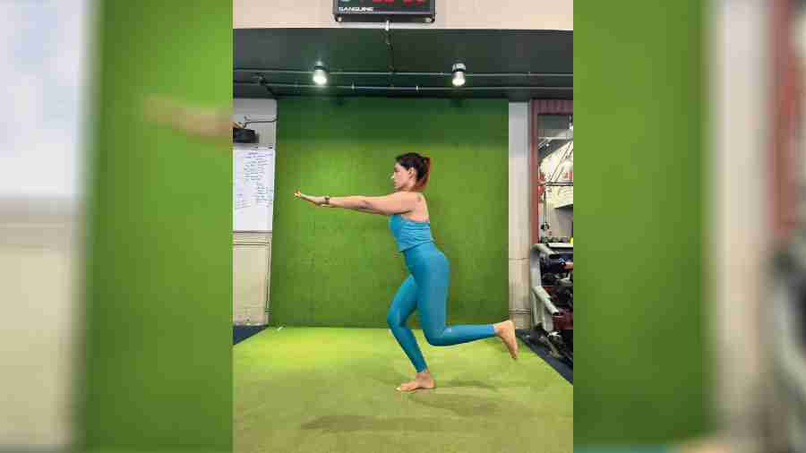
It is increasingly becoming clear in the clinical community that the progression of the initial changes to the structural integrity of a joint can be slowed down
- Remember that it may not be aflare: It may be due to emotional stress, season change or even underlying health conditions.
- Adjust your activity and expectations: When a flare is severe, it may not be possible to get up or move around much. Be kind to yourself and give yourself some flexibility.
- Prepare for your flares: Ask your doctor what medication, topical gels or NSAIDS you can use when you get a flare.
- Stay active on the days when you don’t have flares: Follow your regular corrective and strength/ mobility exercise programme as it will help to reduce the frequency of flares.
- Prioritise good sleep: Our bodies heal during sleep. Getting quality sleep is one of the most important things you can do when you have a condition such as AS/RA.
- Build a support network: Work with health coaches and therapists who actually understand your condition and help tide you over the bad times.
This columnist has been suffering from Ankylosing spondylitis, a form of debilitating auto-immune dysfunction, for the last 35 years or so. This disease involves inflammation inthe hips, sacroiliac joints and spine. Over time, the patient develops a “bamboo spine” and finds mobility very difficult. The author has been taking prescription NSAIDS (nonsteroidal anti-inflammatory drugs) throughout this period and immunosuppressant injections off and on over the last five years or so. But the mainstay of his treatment has been participating in aggressive rehabilitative and corrective exercises. He has successfully slowed down the advent of his disease through exercise and lifestyle modifications.
Exercise and arthritis
It is increasingly becoming clear in the clinical community that the progression of the initial changes to the structural integrity of a joint can be “slowed down” through a rehabilitation programme involving exercises that target strength and flexibility of the muscles and other connective tissue surrounding the joint.
A structured and well-designed exercise programme will slow down the progression of the degenerative changes already existing in the joint.
Programme guidelines and considerations
- Patients should always perform an adequate warm-up session (10 minutes) to ensure joint lubrication and increased elasticity of tissues. They should start with light aerobic exercise to increase systemic blood flow and body temperature.
- They should perform activation exercises to target specific areas (knees, hips) focusing on the full range of motion (ROM).
- Dynamic flexibility exercises should be performed to maintain elasticity and further increase lubrication.
From personal experience, I find that static stretching tends to cool the body down while dynamic movements keep it warm and moving. In my next column I will describe in detail exercises for tackling knee OA.
By increasing muscular strength and endurance, enhancing the stability of the joints, improving a range of motion of the joints and reducing passive tension of the soft tissue around the joints, a patient can improve the quality of their life, maintain normal function and prevent deconditioning.
One of the secondary outcomes of OA is the development of other diseases such as coronary artery disease, diabetes and hypertension. As physical activity becomes too painful to attempt, cardiovascular function declines and the subject becomes sedentary.
Unfortunately, exercise is not a cure for OA, but maintaining a regular exercise programme can reduce pain and rate of decline in functional capacity (Ettinger et al, 1997). No evidence exists that properly programmed and managed exercise will increase the rate of joint degeneration, as measured by joint-space narrowing or pain scores (Mickesky et al,2006).
Each exercise incorporated into the rehabilitative programme for the individual with OA must be evaluated for the amount of force it places on the vulnerable joint, because although the individual may be able to perform the exercise without any pain, this is no guarantee that the exercise is not doing more harm than good over an extended period of time.
A big factor for the increase in the prevalence of OA in India in the last 30 years or so has been dietary and lifestyle habits. Indians are also living longer and developing arthritic changes in hips and/or knees that had never experienced acute trauma in the past. Being overweight or obese also raises the chance of developing arthritis by many folds. There are many people who will develop OA with no identifiable underlying causes; a vast majority of OA is secondary to trauma and or/obesity. Therefore, exercise programmes for individuals with OA must keep forces on the osteoarthritic joint to a minimum, as patients strive to regain the flexibility and strength necessary for a joint to function normally.
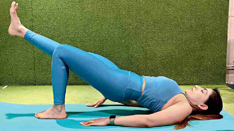
Maintaining a regular exercise programme can reduce pain and rate of decline in functional capacity
Because it is relatively silent in nature in the early stages, OA is comparable to hypertension in that they are both diseases that continue to worsen without telltale signs. These changes tend to worsen over time and have dramatic effects on an individual’s health and lifestyle activities if he/ she fails to recognise them in the early stages. The diagnostic criterion that is widely used to identify individuals with OA of the knee as outlined by the Agency for Healthcare Research and Quality are joint pains plus five of the following criterion (Samson et al., 2007):
- Over 50 years of age
- Less than 30 minutes of morning joint stiffness
- Crepitus
- Bony tenderness
- Bony enlargement
- No palpable warmth of synovium
- Erythrocyte Sedimentation Rate (ESR)< 40 mm/hr
- Rheumatoid factor< 1:40
- Non-inflammatory synovial fluid
Sample exercises
Free-weight and body-weight exercises are most preferred for subjects with OA as they allow for the development of neuromuscular control and conditioning for antagonists and synergists to help control and stabilise the joint.
Sometimes it is a big challenge to increase quadriceps strength in subjects without increasing the risk of joint degeneration. The open chain 30 degrees knee extension exercise is an effective means of doing just that. This exercise allows for the strengthening of the knee extensors but by performing only the last 30 degrees, the subject avoids the high compression forces of the full ROM knee extension.
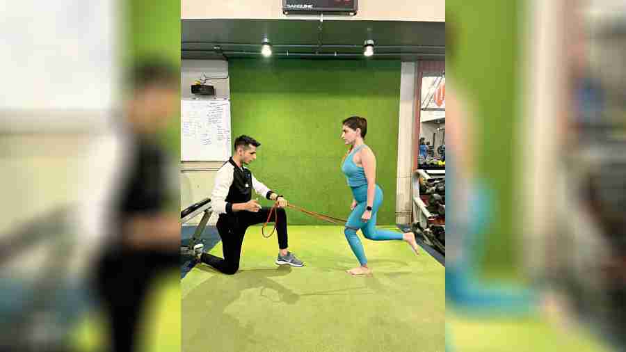
The next progression would be teaching the client to perform closed-chain terminal knee extensions. This exercise allows for the development of the quadriceps but also hip extensors and abductors in a stabilising capacity.
As the client progresses further,the addition of bodyweight exercises to condition the lower body is recommended. One such exercise is the single-legged squat. These exercises place great demand on the client’s ability to control and stabilise the pelvis and femur. Many clients lack the hip strength required to maintain proper pelvic alignment. Two common errors are allowing the knees to move laterally during the eccentric phase of the exercise (descent) and medially (inwards) during the concentric phase (ascent) of the exercise. Note: individuals displaying medialmotion of the knees often have poorly functioning external rotators and extensors (gluteal group) or tight adductors.
The one-legged hip bridge is a great way to resolve this problem. Performing the hip bridge strengthens the gluteal and hamstring group of muscles and helps to stabilise the hip — this ensures better tracking of the patella (knee cap) during lower body movement.

Ranadeep Moitra is a strength and conditioning specialist and corrective eexercise coach
